REAL-TIME IN VIVO CELLULAR IMAGING
Cellvizio lets you see precisely what’s occurring in the human body, at the cellular level in real time.
Know precisely where to target biopsies. Examine cells from inside a nodule. Discover previously undetected lesions. See the immediate impact of a drug on cells.
It’s visibility on a micro level, with macro potential to transform patient management and care.
ACCELERATE DIAGNOSIS. INFORM TREATMENT.
IMPROVE THERAPY.
Used globally across a wide range of medical specialties, including gastroenterology, pulmonology, urology, and drug development, Cellvizio is transforming the way physicians diagnose and treat patients.

Monitor
The progression of disease over time
Detect and monitor cellular changes in vivo which cannot be observed any other way (GERD/Barrett’s Esophagus, Gastric Diseases, COVID-19, Colorectal Lesions)

Assess
Point-in-time reactions, as they happen in real time
Observe and understand cause-and-effect responses to determine the optimal course of action (IBS Food Allergies, Inflammatory Bowel Diseases)

Classify
Indeterminate areas of concern
Confidently characterize areas of interest in areas where conventional disease screening returns ambiguous results (Pancreatic Cysts, Lung Nodules, Bilio-Pancreatic Strictures)

Guide
Surgical interventions
Plan, intervene and treat more precisely with image-guided surgery (Urology)
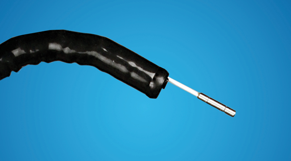
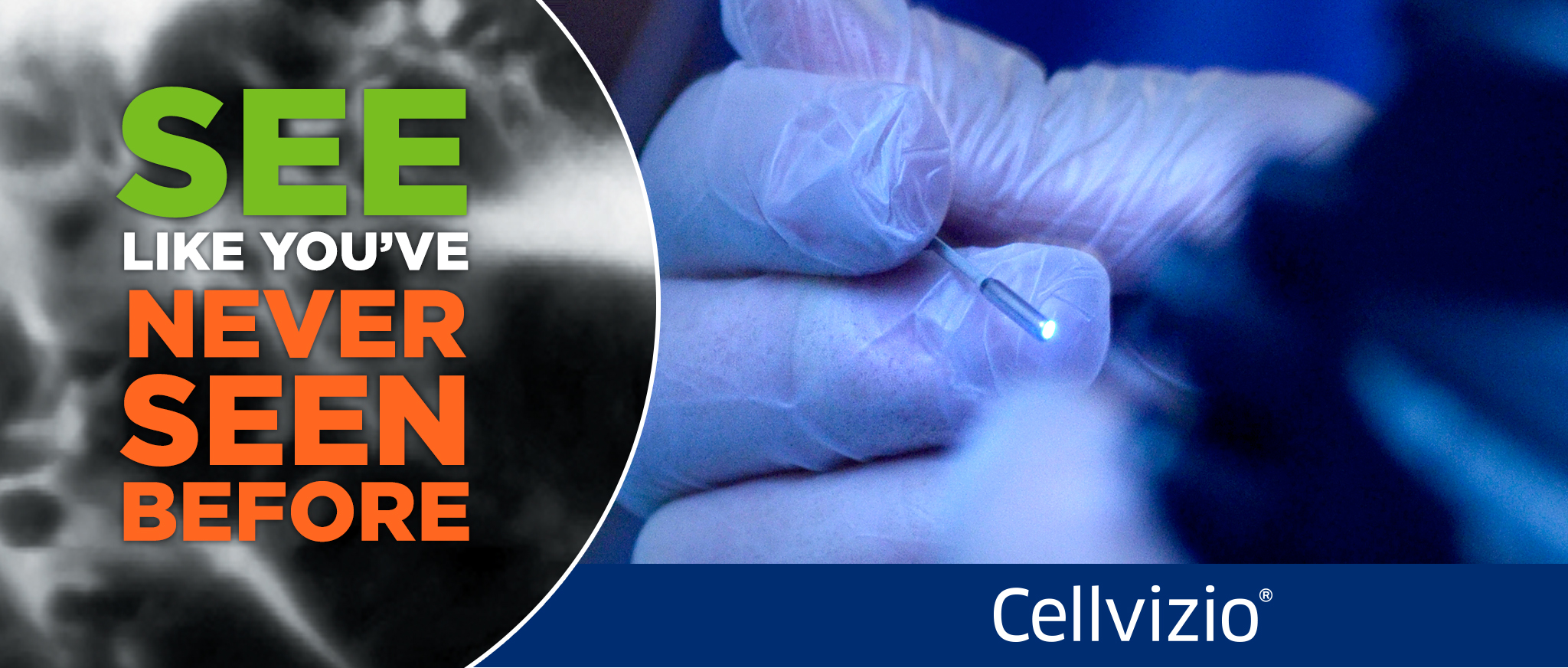
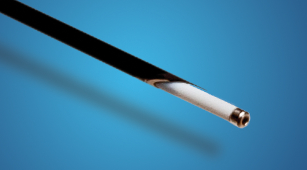
pCLE (Probe/Catheter)
nCLE (Through the Needle)
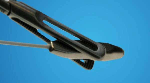
pCLE (Laparoscopic/Robotic)
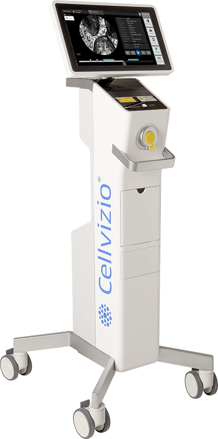
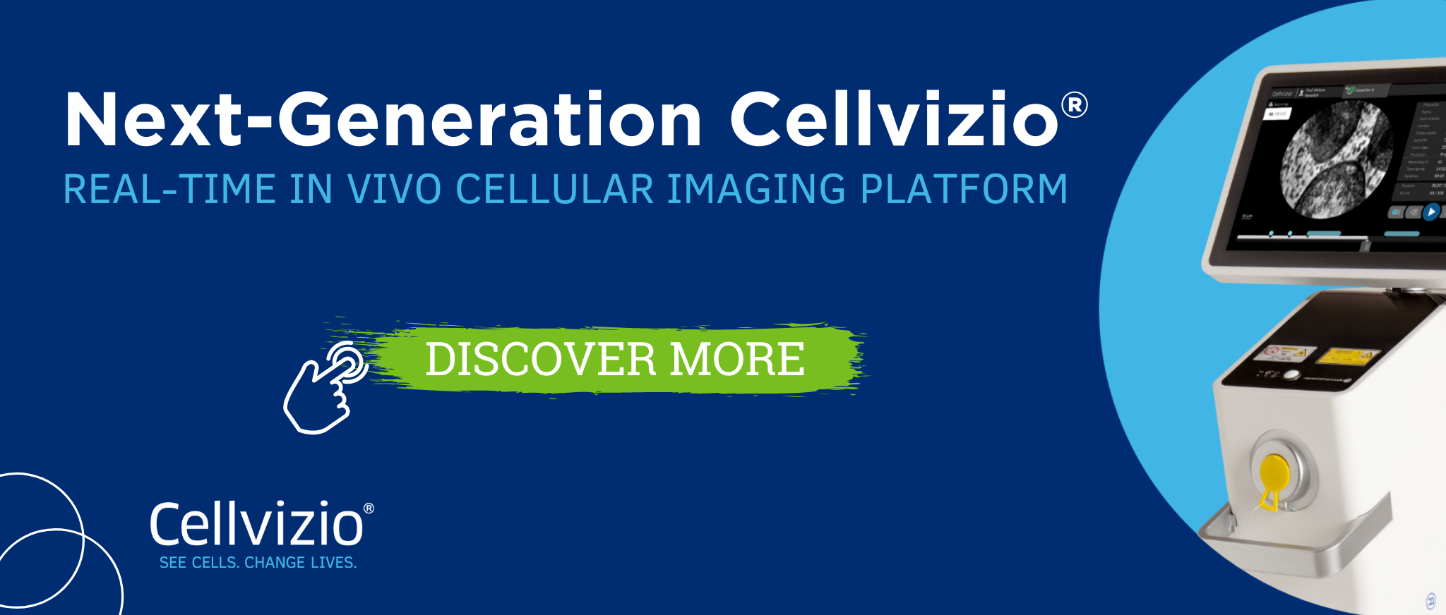
The cellvizio platform
The Cellvizio real time in vivo cellular imaging platform uses Confocal Laser Endomicroscopy (CLE) advanced imaging technology to deliver cellular visualization virtually anywhere in the human body. The system is intuitive and easy to operate, using Confocal Miniprobes™ inserted into the working channel of standard endoscopes.
Learn moreGERD/BARRETT’S ESOPHAGUS MONITORING

Cellvizio® makes it possible for us to: rule-in or rule-out the presence of pre-cancerous cells (...) perform improved surgical intervention (...) monitor for the presence of cancerous cells after the procedure.
Dr. J. Burnette, Coliseum Northside Hospital

