

Mauna Kea Technologies ← InvestorsFrançais

Financial communication & shareholder insight (Euronext Growth: ALMKT)

Pancreatic cysts: seeing at the cellular level to inform diagnosis

A pancreatic cystic lesion, often simply called a cyst, is usually found by chance. Is it dangerous? With ultrasound alone, the physician cannot always tell. Before comparing the images, here is where we are looking, in three steps.

The wall of a serous cyst, filmed live by Cellvizio
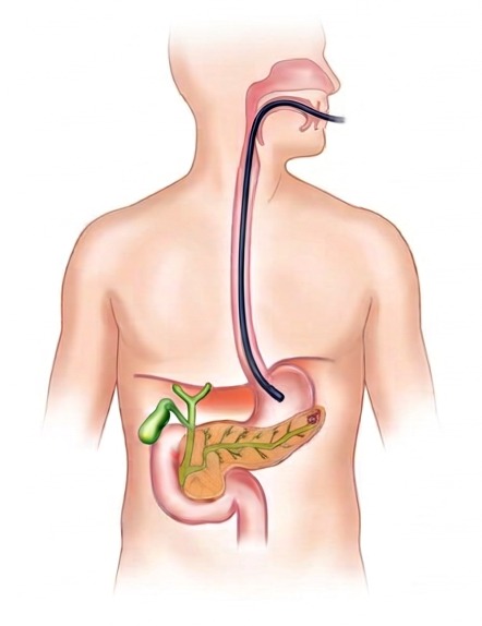
  1. MouthEsophagusUltrasound scopeStomachCystPancreas

    The pancreas is hidden behind the stomach

    The physician reaches it endoscopically: an ultrasound scope goes through the mouth down to the stomach.

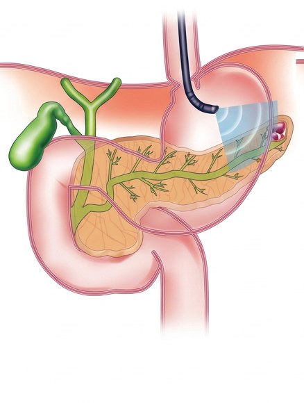
  2. Ultrasound scope Ultrasound Ultrasound image Cyst Pancreas

    The ultrasound scope looks through the wall

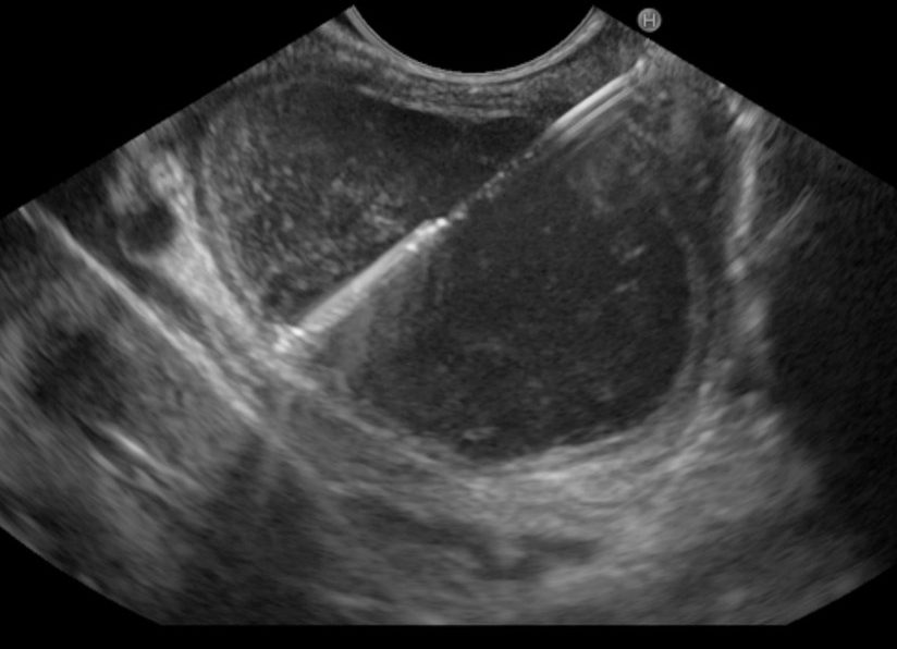
    From the stomach, it sends ultrasound (blue area) toward the pancreas and shows the cyst. This is the ultrasound image in the inset, and the left-hand image of the slider.

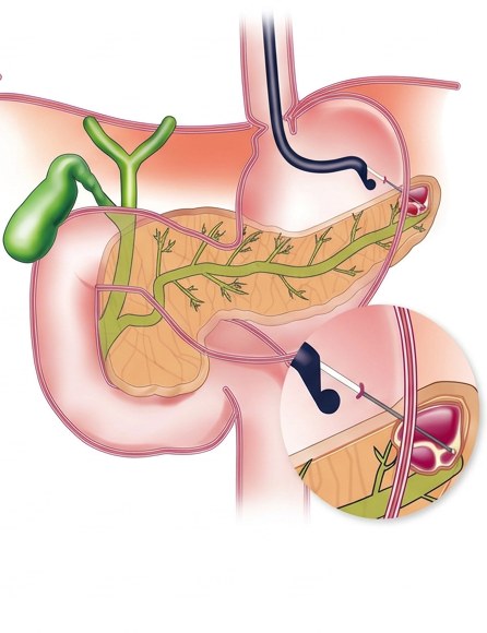
  3. Needle Close-up of the puncture Cellvizio image Cellvizio probe

    The Cellvizio probe films the cyst wall

    A fine needle crosses the wall into the cyst (close-up). The Cellvizio probe is passed through it and touches the wall: the inset shows what it films, the right-hand image of the slider.

Three instruments nested like Russian dolls

The 0.85 mm Cellvizio probe passes through a needle about 1 mm wide, which itself slides through the channel of the ultrasound scope. That is how a microscope reaches the cyst wall, endoscopically.

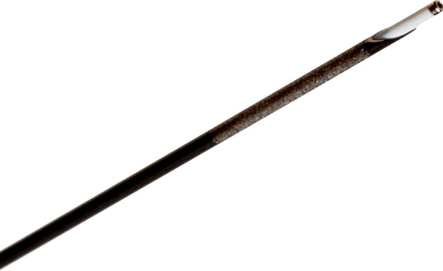
Photo of the Cellvizio probe coming out of the tip of a 19 G needle
Photo: the Cellvizio probe extends from the needle tip
Channel ≈ 3.7 mm Needle ≈ 1.1 mm Probe 0.85 mm Hair ≈ 0.07 mm Cross-section, to scale
Endoscopic ultrasound of a pancreatic cystic lesion, with the aspiration needle
1

Marker:
Drag the slider, or choose a marker from 1 to 5. Marker 3 takes you inside the probe. Illustrative images from two separate exams

Benign or at risk? What the physician looks for

It is the appearance of the wall, as seen by Cellvizio, that guides the diagnosis.

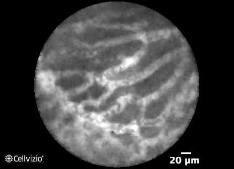
Cellvizio image of a serous cyst: network of vessels
Serous cyst, benignA network of tiny vessels
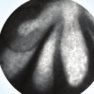
Cellvizio image of an IPMN: papillae
Mucinous cyst (IPMN), at risk of cancerPapillae, finger-like folds

Features described in the literature, to be interpreted by the physician within the complete workup: Napoléon B. et al., Surgical Endoscopy 2016. Cellvizio images from the AQ-Flex 19 brochure, Mauna Kea Technologies.

The medical problem

Pancreatic cystic lesions are often found by chance, on a scan done for another reason. Most are benign; some can progress to cancer.

When standard tests cannot settle the question, some patients undergo surgery as a precaution although their cyst was harmless, while other high-risk cysts are not identified in time.

With Cellvizio, the physician slides a 0.85 mm optical probe through the aspiration needle and looks directly at the cyst wall, at the cellular level.

71%

accuracy of standard cyst fluid analysis (CEA and cytology) in distinguishing mucinous from non-mucinous cysts (INDEX study, n = 144).

60%

of patients with a benign serous cyst undergo unnecessary surgery because of diagnostic uncertainty (Jais et al., Gut 2016, n = 2,622).

40–78%

of surgically removed cysts show neither cancer nor high-grade dysplasia, or nearly 1,850 unnecessary surgeries a year in the United States (Sharib et al., Surgery 2020).

28–37%

postoperative complication rate (morbidity) after pancreatic resection (Krishna Lab, Ohio State University; Jais et al., Gut 2016).

Sources: INDEX study, Krishna et al., Clinical Gastroenterology and Hepatology 2020; Jais et al., Gut 2016; Sharib et al., Surgery 2020; Krishna Lab, The Ohio State University.

What the data show

Accuracy in distinguishing mucinous cysts, at risk of cancer, from the others

Fluid analysis and cytology71%
Cellvizio, during endoscopic ultrasound97%

Prospective single-center INDEX study (Krishna et al., Clinical Gastroenterology and Hepatology 2020, n = 144, Ohio State University). The prospective multicenter CONTACT 2 study (Napoléon et al., Endoscopy 2018, n = 206) reports 100% specificity for serous cysts.

Share of operations deemed inappropriate at Ohio State University, 455 cysts, 2014 to 2024

Before multimodal diagnostics86%
After adopting multimodal diagnostics including Cellvizio6%

Ten-year retrospective observational study (2014–2024) of 455 cysts evaluated at Ohio State University, comparing the periods before and after adoption of a multimodal diagnostic approach including Cellvizio EUS-nCLE (presented by Dr. S. Krishna at DDW 2026). The decline came with no increase in missed resections. The Krishna Lab

Clinical illustration of the care pathway

Documented case — UCLH

Linda B.
“It felt like my life was on hold.”
Linda B., 41, United Kingdom

A CT scan reveals a cyst on the pancreas of Linda B., 41.

At University College London Hospital, Dr. Stephen Pereira examines her as part of a clinical study. Cellvizio endomicroscopic imaging revealed the characteristic superficial vascular network, orienting the diagnosis toward a benign serous cyst and allowing the medical team to rule out major surgical resection.

“It gave me peace of mind,” she says.

Read the full case

This case describes an individual patient journey, documented as part of a clinical study in the United Kingdom. It does not predict the outcome of an exam for any other patient.

Why it matters

Pancreatic cysts are our leading clinical pillar in the United States. Four factors make it a significant market.

Common cysts

3–10%

of adults have an incidentally discovered pancreatic cyst, from 2.6% on CT across all ages up to 8.7% between ages 80 and 89.

An addressable U.S. market

~100,000

patients a year who could benefit from Cellvizio in the United States, our largest market (67% of revenue in the first nine months of 2026).

A validated procedure

CLIMB · ESGE

by the largest U.S. study (CLIMB: 526 patients, 14 centers, ASGE Presidential Plenary at DDW 2026) and included in the European ESGE technical guidelines.

A coded, billable procedure

CPT 43252

An established Category I procedure code allows U.S. facilities to bill for the exam under payers’ fee schedules. Each exam uses a probe, a source of recurring revenue.

Sources: corporate presentation, October 2025, and Laffan et al., AJR 2008; Company estimate, 2025 annual report; nine-month sales press release, October 20, 2026; Cellvizio reimbursement fact sheet, United States.

Learn more

The clinical studies online

The Krishna Lab for Pancreatic Endoscopy website

Dr. Somashekar Krishna’s laboratory at The Ohio State University Wexner Medical Center focuses on the early detection of pancreatic lesions and their organ-preserving endoscopic treatment. Its website brings together in one place its research programs, clinical trials, publications and the international consortium it leads: the best entry point to follow studies of Cellvizio in pancreatic cystic lesions.

This work reaches well beyond one hospital. Each year at DDW, Dr. Krishna convenes the DDW Pancreatic Cyst Consortium: born as the investigators’ meeting of a multicenter Cellvizio study, it drew 123 registrants from more than 12 countries in 2026.

Listen: Dr. Anjuli Luthra on endomicroscopy

In the episode “Earlier, More Accurate Detection of Pancreatic Cancer,” Dr. Anjuli Luthra, co-author of several studies with Dr. Krishna’s team, explains how real-time microscopic imaging helps physicians decide between surveillance, intervention and surgery.

Receive our next short formats and financial communications